
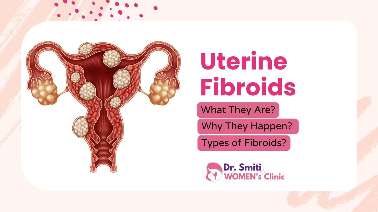
Hearing the words “there is a growth in your uterus” can be frightening. The good news is that in most cases, that growth is a uterine fibroid, a non-cancerous lump that is far more common than most women realise. Many women live with fibroids for years without knowing, and are surprised to find them during a routine ultrasound.
Still, questions come up quickly. What exactly is a fibroid? Why did it develop in my body? Are all fibroids the same? This guide answers these questions in simple language, so you can understand your diagnosis and talk to your gynecologist with confidence.
Quick answer: Uterine fibroids are benign (non-cancerous) growths made of muscle and fibrous tissue in or around the uterus. They develop mainly because of hormonal and genetic factors, and they are classified into types such as intramural, subserosal, submucosal, pedunculated and cervical, depending on where they grow.
What Are Uterine Fibroids?
Uterine fibroids, also called leiomyomas or myomas, are solid growths that develop from the smooth muscle cells of the uterus. Think of them as dense knots of muscle and fibrous tissue that form in the uterine wall.
Key facts at a glance:
- They are benign. Fibroids are not cancer, and having them does not mean you will develop uterine cancer.
- Size varies widely. A fibroid can be as small as a pea or as large as a melon, and in some women it grows big enough to make the abdomen look enlarged.
- Number varies. You may have a single fibroid or several at once.
- They are very common. Fibroids are one of the most frequent gynecological conditions, particularly between the ages of 30 and 50.
- They respond to hormones. Because they depend on estrogen and progesterone, fibroids usually stop growing and often shrink after menopause.
Many women with fibroids never have symptoms. Others notice changes in their periods or pelvic comfort, which is when a gynecology consultation and an ultrasound are advised.
Why Do Uterine Fibroids Occur?
The exact cause of fibroids is still not fully understood. Researchers believe that no single factor is responsible. Instead, a combination of influences works together to trigger a single muscle cell in the uterus to multiply abnormally and form a fibroid.
1. Hormones: estrogen and progesterone
These two female hormones prepare the uterine lining for pregnancy every month. They also appear to stimulate fibroid growth. This is why fibroids develop during the reproductive years, grow when hormone levels are high (for example during pregnancy), and shrink after menopause when hormone levels fall.
2. Genetic changes
Many fibroids carry genetic changes that are different from those in normal uterine muscle cells. Fibroids also tend to run in families. If your mother or sister has had fibroids, your own risk is higher, and you may develop them at a younger age.
3. Growth factors
The body produces natural substances, such as insulin-like growth factor, that help tissues repair and grow. These substances may encourage fibroid cells to multiply.
4. Extracellular matrix (ECM)
Fibroids contain more “cement-like” ECM material than normal muscle. This makes them firm and rubbery, and the matrix also stores growth factors that can further support fibroid growth.
Risk factors that increase the chance of fibroids
- Age: Most common between 30 and 50 years
- Family history: A close relative with fibroids
- Early periods: Starting menstruation before age 11 or 12
- Obesity: Excess body fat can raise estrogen levels
- Never having been pregnant: Pregnancy appears to have a slightly protective effect
- Vitamin D deficiency: Linked in studies, though a direct cause is not proven
- Diet: High intake of red meat and low intake of fruit and vegetables has been associated with higher risk
Important: Fibroids are not caused by anything you did wrong. They are not linked to hygiene, a particular food, or stress alone, and they cannot be “caught” from another person.
Types of Uterine Fibroids
Fibroids are classified by where they grow in relation to the layers of the uterus. The type is important because it influences the kind of changes you may notice and the approach your doctor chooses.
1. Intramural fibroids
- Location: Inside the muscular wall (myometrium) of the uterus
- How common: The most common type
- What to know: As they grow, they can stretch the uterine wall and make the uterus larger. They are often linked with heavy or prolonged periods, a feeling of pelvic pressure, and a bulky lower abdomen. Multiple intramural fibroids can change the shape of the uterus.
2. Subserosal fibroids
- Location: On the outer surface of the uterus, beneath the thin outer lining (serosa)
- What to know: They grow outward into the pelvic cavity and can become quite large without affecting the uterine lining. Because of this, periods may stay fairly normal, but a large subserosal fibroid can press on the bladder or bowel, leading to frequent urination, constipation or back pain.
3. Submucosal fibroids
- Location: Just under the inner lining of the uterus (endometrium), bulging into the uterine cavity
- How common: The least common among the three main types
- What to know: Even a small submucosal fibroid can cause significant heavy bleeding because it sits directly against the lining. Since they alter the uterine cavity, they are the type most associated with difficulty in conceiving, implantation problems and miscarriage.
4. Pedunculated fibroids
- Location: Attached to the uterus by a thin stalk (peduncle)
- Two forms: Pedunculated subserosal (growing outward into the pelvis) and pedunculated submucosal (growing inward into the uterine cavity)
- What to know: The stalk can sometimes twist, cutting off the fibroid’s blood supply. This causes sudden, severe pelvic pain and needs urgent medical attention. Because they are mobile, they can feel like a separate pelvic lump.
5. Cervical fibroids
- Location: In the cervix, the lower neck of the uterus
- How common: Rare
- What to know: Depending on their size, they may cause bleeding between periods, vaginal discharge, or pressure symptoms, and occasionally interfere with the passage of the birth canal.
Other terms you may hear
- Broad ligament fibroids: Grow in the ligament that supports the uterus.
- Parasitic fibroids: Rare fibroids that detach from the uterus and gain a blood supply from another organ.
- FIGO classification: Gynecologists use a numbered system (types 0 to 8) to describe fibroid position precisely. For example, types 0 to 2 are submucosal, types 3 to 4 are intramural, and types 5 to 7 are subserosal.
Conclusion
Uterine fibroids are common, non-cancerous growths of the uterine muscle that develop mainly due to hormonal and genetic influences. They vary widely in size, number and position, and understanding whether yours is intramural, subserosal, submucosal, pedunculated or cervical helps explain what your body may be experiencing and what your doctor needs to look at.
Fibroids are not a reason for panic, and they are nothing you caused. A simple ultrasound and an expert opinion can give you clarity on the type, size and number of fibroids, and whether any action is needed at all. If you have heavy periods, pelvic pressure, or difficulty conceiving, do not ignore it.
Frequently Asked Questions (FAQs)
Which type of fibroid is the most common?
Intramural fibroids, which grow within the muscular wall of the uterus, are the most common type.
Which type of fibroid is most likely to affect fertility?
Submucosal fibroids, because they bulge into the uterine cavity and can interfere with implantation.
Are uterine fibroids cancerous?
No. Fibroids are almost always benign. Cancer in the uterine muscle (leiomyosarcoma) is very rare, and fibroids do not usually turn into cancer.
Can fibroids grow back or disappear on their own?
Fibroids often shrink after menopause because hormone levels drop, but they rarely disappear on their own before that.
At what age do fibroids usually develop?
Most commonly between 30 and 50 years, during the reproductive years when estrogen levels are high.


